23 Pairs of Chromosomes in the Nucleus Containing Hereditary Material :cell Grows and Develops Again
Theme 5: How Do We Command Our Fertility?
5.2 Meiosis and Gametogenesis
Sexual reproduction requiresfertilization, a spousal relationship of two cells from two private organisms. If those ii cells each comprise one set of chromosomes, then the resulting cell contains 2 sets of chromosomes. The number of sets of chromosomes in a cell is called its ploidy level. Haploid cells contain ane gear up of chromosomes. Cells containing two sets of chromosomes are called diploid. If the reproductive bicycle is to continue, the diploid cell must somehow reduce its number of chromosome sets before fertilization can occur once again, or at that place will be a continual doubling in the number of chromosome sets in every generation. Then, in addition to fertilization, sexual reproduction includes a nuclear partition, known as meiosis, that reduces the number of chromosome sets. Meiosis occurs during the process of gametogenesis, which is the production of gametes (oocytes and sperm.)
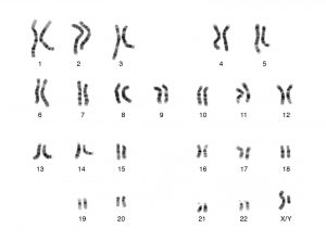
Figure 1 shows a karyotype of a human male person. A karyotype is an image produced by arranging images of each chromosome in a cell into systematic pairs. As you can see, there are 23 full pairs of chromosomes, and 22 of them contain matched pairs of the same size with similar banding patterns, indicating that the cells in each pair contain the same genes. Each of these 22 pairs are called homologous pairs, and the pairs are numbered in society of size, with the 2 copies of chromosome 1 being the largest. The final pair of chromosomes are not matched in size or banding patterns; these are the sex chromosomes and are called the Ten chromosome and the Y chromosome. In most homo cells, there are 22 matched pairs of chromosomes and 1 pair of sex chromosomes. About all males have one X sex chromosome and 1 Y sex chromosome, and near all females have 2 X sex chromosomes, in each cell. Gametes are unique in that they accept half of the chromosomes of other cells: only 1 sex chromosome and only 22 other chromosomes, non 22 pairs.
Most animals and plants are diploid, containing two sets of chromosomes; in eachsomatic cell (the nonreproductive cells of a multicellular organism), the nucleus contains two copies of each chromosome that are referred to as homologous chromosomes. Somatic cells are sometimes referred to every bit "body" cells. Homologous chromosomes are matched pairs containing genes for the aforementioned traits in identical locations along their length. Diploid organisms inherit one copy of each homologous chromosome from each parent; all together, they are considered a full set up of chromosomes. In animals, haploid cells containing a unmarried re-create of each homologous chromosome are establish only within gametes. Gametes fuse with another haploid gamete to produce a diploid cell.
The nuclear division that forms haploid cells, which is called meiosis, is related to mitosis. As you take learned, mitosis is role of a cell reproduction bike that results in identical girl nuclei that are also genetically identical to the original parent nucleus. In mitosis, both the parent and the daughter nuclei incorporate the same number of chromosome sets. Meiosis employs many of the aforementioned mechanisms as mitosis. However, the starting nucleus is always diploid and the nuclei that upshot at the terminate of a meiotic cell sectionalization are haploid. To accomplish the reduction in chromosome number, meiosis consists of i circular of chromosome duplication and two rounds of nuclear division. Considering there are two rounds of division, the stages are designated with a "I" or "Ii." Thus, meiosis Iis the beginning round of meiotic division. Meiosis I reduces the number of chromosome sets from two to one. The genetic information is also mixed during this division to create unique recombinant chromosomes.Meiosis II, in which the second circular of meiotic segmentation takes place in a mode that is like to mitosis, includes all of the stages of division again. These stages have specific names (Prophase, Metaphase, Anaphase, Telophase), but y'all are not required to know the specific names of the stages for this form.
*
Interphase
Meiosis is preceded past a phase called interphase. In this stage, the DNA of the chromosomes is replicated, so that each prison cell contains two copies of each chromatid. This means that in a human being prison cell, both copies of chromosome 1 are copied to produce 4 chromatids, both copies of chromosome two are copied to produce four chromatids, and so on. The cell likewise grows, and produces enough of the enzymes and structures required for meiosis during this interphase period.
*
Meiosis I
Early on in meiosis I, the chromosomes tin can be seen clearly microscopically. As the nuclear envelope begins to intermission down, the proteins associated with homologous chromosomes bring the pair close to each other. The homologous chromosomes now become arranged in the center of the jail cell, with the ends of each pair of homologous chromosomes facing opposite poles. The orientation of each pair of homologous chromosomes at the centre of the cell is random.
This randomness, called contained assortment, is the physical basis for the generation of the 2nd form of genetic variation in offspring. Consider that the homologous chromosomes of a human are originally inherited as two divide sets, one from each parent. One set of 23 chromosomes is present in the egg donated by the mother. The father provides the other prepare of 23 chromosomes in the sperm that fertilizes the egg. These pairs line up at the midway bespeak betwixt the 2 poles of the jail cell, and their arrangement in regard to the two poles is random. Whatever maternally inherited chromosome may face up either pole. Any paternally inherited chromosome may likewise face either pole. The orientation of each pair is independent of the orientation of the other 22 pairs.
In each cell that undergoes meiosis, the organisation of the chromosomes is different. There are 2 possibilities for orientation (for each pair); thus, the possible number of alignments equals 2 northward wherenorth is the number of chromosomes per set. Humans have 23 chromosome pairs, which results in over 8 million (223) possibilities. Other mechanisms not discussed in this class can increase the variation in each prison cell produced besides. Given both independent assortment and these other mechanisms, it is highly unlikely that any two haploid cells resulting from meiosis will have the same genetic composition.
Next, protein fibers in the jail cell pull the linked chromosomes apart. The fibers pull the chromosome to the opposite poles of the cell. At each pole, at that place is simply one fellow member of each pair of the homologous chromosomes, so just one total set of the chromosomes is nowadays. This is why the cells are considered haploid—there is only one chromosome prepare, even though there are duplicate copies of the gear up because each homolog still consists of 2 "sis: chromatids that are still attached to each other. Cytokinesis, where the cell membrane pinches off in the middle of the cell to separate it into ii, now occurs, splitting each jail cell into two cells containing one full ready of chromosomes.
Concept in Activity
Review the process of meiosis, observing how chromosomes align and migrate, at this site.
*
Meiosis II
In meiosis II, the connected sister chromatids remaining in the haploid cells from meiosis I will exist dissever to class four haploid cells. The two cells produced in meiosis I go through the events of meiosis II in synchrony. Overall, meiosis Ii resembles the mitotic sectionalisation of a haploid cell.
First, the nuclear envelope breaks downwards and the chromosomes are clearly visible under a microscope. The sister chromatids then line upwards at the centre of the jail cell. Protein fibers pull one of each pair of sisters toward the poles of the cell.The chromosomes arrive at reverse poles.. Nuclear envelopes grade around the chromosomes. Cytokinesis separates the 2 cells into four genetically unique haploid cells. At this point, the nuclei in the newly produced cells are both haploid and have but one copy of the unmarried set of chromosomes. The cells produced are genetically unique considering of the random assortment of paternal and maternal homologs and because of other sources of variation non discussed here.
*
Comparing Meiosis and Mitosis
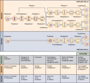
Mitosis and meiosis, which are both forms of segmentation of the nucleus in eukaryotic cells, share some similarities, just also exhibit distinct differences that lead to their very different outcomes. Mitosis is a single nuclear division that results in two nuclei, usually partitioned into 2 new cells. The nuclei resulting from a mitotic division are genetically identical to the original. They take the same number of sets of chromosomes: i in the instance of haploid cells, and 2 in the example of diploid cells. On the other hand, meiosis is ii nuclear divisions that effect in four nuclei, usually partitioned into iv new cells. The nuclei resulting from meiosis are never genetically identical, and they contain i chromosome set only—this is half the number of the original jail cell, which was diploid ( Figure 2 ).
The differences in the outcomes of meiosis and mitosis occur because of differences in the behavior of the chromosomes during each process. Most of these differences in the processes occur in meiosis I, which is a very dissimilar nuclear sectionalization than mitosis. In meiosis I, the homologous chromosome pairs become associated with each other, are bound together, feel chiasmata and crossover between sister chromatids, and line up forth the metaphase plate in tetrads with spindle fibers from opposite spindle poles fastened to each kinetochore of a homolog in a tetrad. All of these events occur just in meiosis I, never in mitosis.
Homologous chromosomes movement to opposite poles during meiosis I so the number of sets of chromosomes in each nucleus-to-be is reduced from two to i. For this reason, meiosis I is referred to as areduction division. There is no such reduction in ploidy level in mitosis.
Meiosis Two is much more analogous to a mitotic division. In this case, duplicated chromosomes (but one set of them) line upwards at the center of the cell with divided kinetochores attached to spindle fibers from reverse poles. As in mitotis, one sis chromatid is pulled to one pole and the other sis chromatid is pulled to the other pole during meiosis II. Meiosis II is not a reduction sectionalization because, although there are fewer copies of the genome in the resulting cells, there is still one set of chromosomes, as there was at the finish of meiosis I.
Cells produced by mitosis will function in different parts of the body equally a part of growth or replacing dead or damaged cells, only cells produced past meiosis will but participate in sexual reproduction.
Concept in Activity
For an animation comparing mitosis and meiosis, go to this website.
*
Gametogenesis (Spermatogenesis and Oogenesis)
Gametogenesis, the product of sperm and eggs, includes the process of meiosis to produce haploid cells, and growth and maturation of these cells into oocytes and sperm. The production of sperm is called spermatogenesis and the product of eggs is called oogenesis.
Spermatogenesis
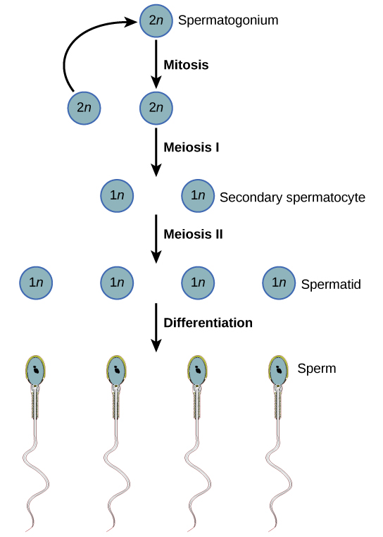
Spermatogenesis, illustrated in Effigy three , occurs in the wall of the seminiferous tubules, with stem cells at the periphery of the tube and the spermatozoa at the lumen of the tube. Immediately under the capsule of the tubule are diploid, undifferentiated cells. These stalk cells, called spermatogonia (atypical: spermatagonium), go through mitosis with one offspring going on to differentiate into a sperm cell and the other giving rise to the next generation of sperm.
Meiosis starts with a jail cell called a primary spermatocyte. At the end of the commencement meiotic partition, a haploid cell is produced chosen a secondary spermatocyte. This cell is haploid and must get through some other meiotic cell division. The cell produced at the cease of meiosis is called a spermatid and when it reaches the lumen of the tubule and grows a flagellum, it is chosen a sperm jail cell. Iv sperm result from each primary spermatocyte that goes through meiosis.
Stem cells are deposited during gestation and are present at birth through the beginning of adolescence, simply in an inactive state. During adolescence, gonadotropic hormones from the inductive pituitary cause the activation of these cells and the production of viable sperm. This continues into old historic period.
Link to Learning
Visit this site to see the process of spermatogenesis.
Oogenesis
Oogenesis, illustrated in Figure 4 , occurs in the outermost layers of the ovaries. As with sperm production, oogenesis starts with a germ prison cell, called an oogonium (plural: oogonia), simply this prison cell undergoes mitosis to increase in number, eventually resulting in up to about one to two million cells in the embryo.
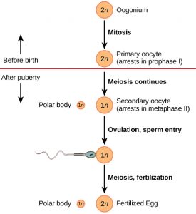
The prison cell starting meiosis is called a principal oocyte, equally shown in Effigy iv . This cell will start the meiosis I and exist arrested in its progress very early, in a stage called the starting time prophase phase. At the fourth dimension of nascence, all future oocytes are in the prophase stage; no additional oocytes or precursors are produced after birth. At adolescence, inductive pituitary hormones cause the development of a number of follicles in an ovary. This results in the primary oocyte finishing meiosis I. The cell divides unequally, with virtually of the cellular fabric and organelles going to i cell, chosen a secondary oocyte, and merely one set of chromosomes and a small corporeality of cytoplasm going to the other cell. This second cell is chosen a polar trunk and normally dies. A secondary meiotic arrest occurs, about halfway through the meiosis 2 in a stage called the metaphase 2 stage. At ovulation, this secondary oocyte will be released and travel toward the uterus through the oviduct. If the secondary oocyte is fertilized, the cell continues through meiosis II, producing a second polar body and a fertilized egg containing all 46 chromosomes of a homo existence, half of them coming from the sperm. If the oocyte is not fertilized, notwithstanding, it does non complete meiosis Ii.
Oocyte production begins earlier birth, is arrested during meiosis until puberty, and and so individual cells continue through at each menstrual bicycle. Ane oocyte is produced from each meiotic process, with the extra chromosomes and chromatids going into polar bodies that degenerate and are reabsorbed by the body.
Section Summary
Sexual reproduction in humans requires that diploid individual cells produce haploid cells that can fuse during fertilization to form diploid offspring. The process that results in haploid cells is called meiosis. Meiosis is a serial of events that accommodate and separate chromosomes into daughter cells. During the interphase of meiosis, each chromosome is duplicated. In meiosis, there are two rounds of nuclear division resulting in four nuclei and ordinarily 4 haploid daughter cells, each with half the number of chromosomes as the parent cell. During meiosis, variation in the daughter nuclei is introduced because of random alignment in meiosis I. The cells that are produced past meiosis are genetically unique.
Meiosis and mitosis share similarities, but have singled-out outcomes. Mitotic divisions are unmarried nuclear divisions that produce daughter nuclei that are genetically identical and have the aforementioned number of chromosome sets equally the original cell. Meiotic divisions are 2 nuclear divisions that produce iv daughter nuclei that are genetically different and have one chromosome gear up rather than the two sets the parent cell had. The main differences between the processes occur in the first partition of meiosis. The homologous chromosomes separate into unlike nuclei during meiosis I causing a reduction of ploidy level. The second division of meiosis is much more similar to a mitotic partition.
Important similarities exist between spermatogenesis and oogenesis: during both processes a diploid cell is duplicated a number of times in mitosis to produce precursors that undergo two rounds of meiosis to produce haploid sperm and eggs. Of import differences exist in the timing of these divisions and in the symmetry of divisions. In males, each spermatagonium produces four mature sperm, but in oogenesis each oogonium can produce only 1 fertilized egg.
*
Glossary
- fertilization
- the union of ii haploid cells typically from 2 individual organisms
- karyotype
- systematic arrangement of images of chromosomes into homologous pairs
- meiosis I
- the kickoff round of meiotic cell division; referred to as reduction sectionalisation considering the resulting cells are haploid
- meiosis II
- the second round of meiotic prison cell division post-obit meiosis I; sister chromatids are separated from each other, and the consequence is four unique haploid cells
- oogenesis
- the process of producing haploid eggs
- reduction partitioning
- a nuclear division that produces daughter nuclei each having one-half equally many chromosome sets as the parental nucleus; meiosis I is a reduction sectionalization
- somatic prison cell
- all the cells of a multicellular organism except the gamete-forming cells
- spermatogenesis
- the process of producing haploid sperm
Source: https://open.lib.umn.edu/humanbiology/chapter/5-2-meiosis/
0 Response to "23 Pairs of Chromosomes in the Nucleus Containing Hereditary Material :cell Grows and Develops Again"
Post a Comment